Crouzon syndrome is a congenital genetic disorder characterized by an anomalous fusion or bonding between the bones of the face and the skull.
In normal cases, during the development of the brain, open sutures occurring between the bones permit the normal growth of the skull. However, when the bone sutures close earlier than normal, then the skull tends to form in the direction of the sutures that are still non-fused. Patients of Crouzon syndrome experience earlier than normal fusion of the facial and skull bones, which then results in abnormally shaped face, head, and teeth.
The FGFR2 or fibroblast growth factor receptor genes occurring in chromosome 10, or the FGFR3 genes on chromosome 4 experience a mutation or genetic error which causes Crouzon syndrome.
Crouzon syndrome is an uncommon condition and tends to affect an estimated 1.6 individuals per 100,000 people. They constitute around 4.5 percent of the patients affected by craniosynostosis disorder.
Symptoms of Crouzon syndrome
The following are some of the signs and symptoms of Crouzon syndrome:
- Eye abnormalities
- Improper alignment between the eyes leading to vision problem
- Increased distance or gap between the two eyes
- Shallow eye sockets leading to projection of eyeballs from their respective sockets
- Facial abnormalities
- Low set ears, which can increase the risk to complete or partial loss of hearing
- Lack of normal growth in the mid-face and upper jaw
- Beak like nose formation
- Narrow and tall palate
- A protruding chin
- All the above listed abnormal face and skull features lead to difficulties in breathing
- Pigmented and dark patches on the sides of the mouth, neck, and eye lids
- Other features
- Extreme instances of Crouzon syndrome may occur along with Meniere’s disease.
- Shorter growth of femur bone and the bone of forearm
- Partial syndactyly
- Hands and legs are free from defects
Types of Crouzon syndrome fusions
The varied patterns of fusion associated with Crouzon syndrome are listed below:
- Fusion of metopic suture: It is identified by the formation of a triangular shapedforehead. It is caused due to premature fusion of two frontal bones that restricts transverse growth of the skull and results in its parallel growth, thereby causing the triangular appearance of the skull.
- Fusion of sagittal suture: It causes the formation of narrow and long head, like an inverted boat, due to premature bonding of the two parietal bones
- Fusion of coronal structure: It is caused due to premature fusion of frontal bones with parietal bones, that results in a ‘front to back’ shortened skull diameter, i.e. flat head.
- Fusion of lambdoid and coronal suture: It refers to premature fusion of the skull bones that result in asymmetrical distortion of head, wherein the skull bones get flattened towards one side.
- Fusion of lamdboidal and coronal suture:It refers to premature fusion of parietal and occipital skull bones leading to oxycephalyor the high headed syndrome.
- Fusion of all sutures, results in Kellebalttscheaedel.
Causes
Crouzon syndrome is an inherited autosomal dominant disorder. It is caused due to mutations or errors in the fibroblast growth factor receptor or FGFR2 genes. Less commonly, it is caused due to mutated FGFR3 genes. Defects in any of these genes can result in premature fusion of the bones in the skull.
The genetic mutations can be passed on from any one of the affected parents. Patients carry a fifty percent risk of passing on the genetic anomaly to their child.
Crouzon does not result in defects of the legs and hands and hence is not similar to other kinds of craniosynostosis syndromes. However, abnormalities of the cervical spine are common.
Diagnosis
Apart from the signs and features listed above, diagnosis involves the use of below listed devices and tests to verify the correctness of diagnosis:
- X-ray
- CT scan
- Genetic tests
- Test of sample cells
- Information about a family history of the disease
Crouzon syndrome Treatment
Depending on the type of abnormal fusion, surgery is performed to correct Crouzon syndrome defects.A particular kind of plastic surgery,called craniofacial surgery,is the major remedy fortreating Crouzon syndrome. The procedure is carried out by a plastic surgeon along with the help of neurosurgeons.
As the different fusions involve more than one suture, an open surgery is normally performed on affected patients.The following types of surgeries may be needed:
- For correcting the mid face disorder, plastic surgeons need to move the maxillary bone and the lower orbit forward. It will help correct the eye socketsshape defect.
- Cranial surgery is done for correction of the shallow orbits, Chiari dysfunction, defective ear canal, sleep apnea, respiratory and speech defects, and hydrocephalus condition, etc.
- The surgeon performs plastic surgery to correct deformities of the upper jaw, as well as other defects within the oral cavity like crowding of teeth or hypodontia, bilateral cross bite, and high arched palate
- The orbital cavity is reshaped and the orbits moved forward for realigning the eyes
- Follow up re-correction surgery is also a part of the normal treatment routine of Crouzon syndrome.
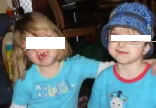
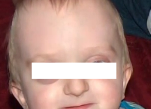
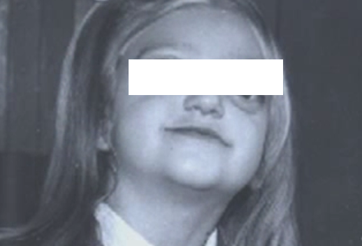
Crouzon syndrome Pictures
