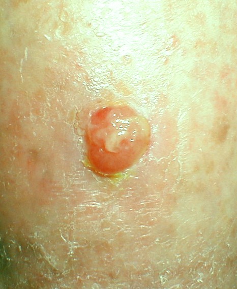
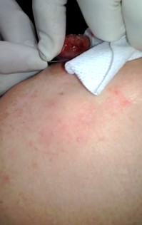
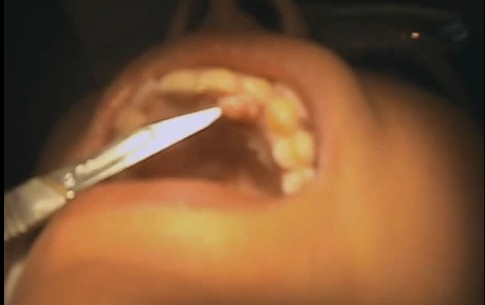
Pyogenic granuloma or pregnancy tumor usually starts as a lesion and then grows rapidly for few weeks. They appear like small, red bumps on the skin. These bumps appear as an overgrowth of tissue due to hormonal factors, irritation and physical trauma. They appear like reddish or yellowish nodule and usually smaller than 2 cms. The tender lumps usually have a smooth surface with moist appearance; but have a crusty or rough surface with lot of bleeding. Bleeding usually takes place because of large number of blood vessels present. Although the symptoms of pyogenic granuloma are seen on people of all ages, they mainly affect children and young adults. Pyogenic granuloma is also called pregnancy tumor because they are commonly found in women who are pregnant, mainly because of the hormonal changes that take place during pregnancy. They are non cancerous and non infectious and can be safely removed.
The name Pyogenic granuloma first came into picture in 1897 by two French surgeons, Poncet and Dor. The duo named this lesion as botryomycosis hominis which was later renamed as pyogenic granuloma.
Causes of Pyogenic granuloma
The causes of Pyogenic granuloma is unknown. However, several factors are considered to play their role in the development of pyogenic granuloma. It is usually caused by trauma, especially on the sites where individuals have suffered recent injuries. Infection, possibly viral infection is also said to be a main cause of these tumors. It is also believed that pregnant women have high risk of being affected by these lumps due to hormonal imbalances. It has been observed that patients who are on systemic retinoids or portease inhibitors also get induced to multiple lesions. Pyogenic granuloma affects young adults and children but pregnant women are more vulnerable to these. Granuloma infects pregnant women in the first three months till seventh month and is seen in anterior nasal septum when further cause frequent nose bleeds. Women have more risk of granuloma than in men.
Symptoms
Pyogenic granuloma usually occurs in arms, face, hands and in the mouth. Lips, tongue and inner cheek are seen as common areas where granuloma can be infected. Anterior areas are more prone to granuloma than posterior areas. The color of the lesions ranges from red or pink to even purple and has a smooth or lobulated head and sometimes bleeds. When the lesions start developing, it appears red in color due to the high number of blood vessels present there. The color may also appear to be brownish red or blue black. The color however, changes to pink when lesions become little old.
Once these lesions get developed, they rapidly grow for few weeks and their size differ from 2mm to 2cm in diameter. Most of the times, a single lesion is observed; but in certain circumstances multiple lesions can also be seen. They bleed so much and may form ulcers and then a crusted sore. Sometimes, oil like substance gets leaked which dampens the surface of the lump. They are painful and if the location of this lesions are in an area which are constantly being disturbed, then they become more painful and causes lot of discomfort to the sufferers.
Diagnosis of Pyogenic granulomas
Pyogenic granulomas are diagnosed clinically by physical examination and medical tests. A physical examination of lesions, its appearance and time course plays a crucial factor in the diagnosis. Doctors may also ask the patients to go for a biopsy to rule out any possibility of cancer. Frequent visits to dermatologists are requires to diagnose the granuloma and to rule out any kind of malignant cancer and melanoma.
Treatment of pyogenic granulomas
The treatment of pyogenic granulomas depends on their size. Most medical professionals leave the small granulomas as it without any treatment. They usually go away on their own and if they don’t disappear after 4 weeks, then they are required to be treated differently. Larger pyogenic granulomas are required to be surgically removed. Most likely, doctors may scrape it off and then cauterize or burn it completely. Doctors may also apply few chemicals like silver nitrate which will drain the blood completely from the infected lesion. The surgery prescribed is minor and few of the granulomas may also be removed by laser treatment. Sometimes, doctor may advise for pulse dye laser for removing the small lesions. No treatment is prescribed to pregnant ladies. Patients suffering from granulomas are advised not to pick them of as this may aggravate the problem.
Complications
Frequent bleeding from the lesions can cause complications. There are possibilities the lesions may grow back after their removal. In some cases, the lesions redevelop in the same area. If proper care is not taken while removing the granulomas, the remaining parts might get spread to blood vessel in the same area.
Pyogenic granuloma pictures


